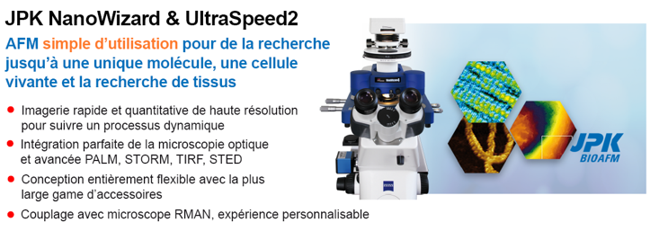
Ateliers Pré-congrès > Atelier BrukerCaractérisation des matériaux aux petites échellesProgramme de l'atelier : 10h00 -10h10 Présentation de la gamme d’instruments Bruker – Jérôme Beaumale Ingénieur France 10h10 – 10h55 - Nanoscale Mechanobiology by AFM – from Single Molecules via Living Cells towards Tissues - TBA Applications Scientist, Bruker Nano Systems and Metrology, Berlin, Germany In recent years Atomic Force Microscopy (AFM) has emerged as a versatile tool no longer limited to materials research. Evolution in design and usability made it a powerful tool for life science research. Here, we present our multipurpose AFM platform allowing comprehensive characterization of biological samples, such as, live cells, tissues and biomaterials at the nanoscale. The nano-mechanical analysis of cells and tissues is increasingly gaining in importance in different fields of cell biology, such as, cancer research and developmental biology. Here, we will show how nanomechanical data can be obtained on biologically relevant samples by different approaches. Using single cell force spectroscopy (SCFS), cell-substrate or cell-cell/tissue interactions can be investigated and quantified down to single protein unbinding events. AFM based SCFS turned into a well-established technique giving insights into the early cell adhesion process. With our Quantitative Imaging (QI™) mode several sample properties, such as the topography, stiffness and adhesion, can be obtained with one measurement at high resolution. Even more complex data like Young´s modulus images or recognition events can be extracted. A variety of biological samples will be discussed to demonstrate the capability and flexibility of QI™. The instrument demonstration will introduce the relevant AFM basics and the workflow from sample preparation to AFM measurements and data analysis. Remote demonstration from Berlin of the NanoWizard4 Atomique Force Microscopy – mechanical study biological object.
11h00- 11h45 Etat de l’art en indentation : des matériaux durs au matériaux biologiques– Michel Fajfrowski Ingénieur application Bruker La nanoindentation est une technique qui est devenue aujourd’hui extrêmement usitée. Après des premiers développements dans les années 1980, elle est devenue une technique industrielle pour certaines applications. Grace aux progrès récents de la micro électronique il est maintenant possible de générer des cartographies de propriétés mécaniques associées à des observation à fort grandissement. De même, de nouvelles gammes de matériaux sont maintenant accessibles, notamment biologique et/ou dépendants du temps. Cette connaissance à nécessité la création de nouveaux modèles qui seront brièvement présentés. Il ne faut néanmoins pas oublier que les fondamentaux restent inchangés et qu’une analyse rigoureuse est nécessaire pour éviter de fausses interprétations. C’est pourquoi cette présentation se focalisera également sur les paramètres critiques à valider. Une procédure sera proposée pour permettre à des utilisateurs non initiés à éviter des erreurs et valider leurs données. … Démonstration avec l’indenteur Bruker TS-77 sur divers matériaux.
|
| Personnes connectées : 24 | Flux RSS | Vie privée |

|


